 From the Coloradoan.com
Colorado - Barney was a Great Pyrenees who was born with a bow-shaped leg bone that caused a dislocated knee cap. It started when he was a pup at just a month old, where the pain prevented Barney from putting weight on his right back leg.
But less than a year ago a revolutionary type of orthopedic surgery changed Barney's quality of life.
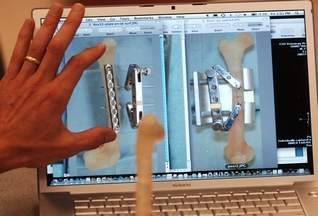
In September 2007, Dr. Ross Palmer, an associate professor at the Colorado State University veterinary teaching hospital, using new technology more advanced than X-rays, created 3-D images of Barney's femurs. The CT scan used to generate the images creates more accurate measurements and visual models of the bone.
ProtoMed Inc., an Arvada-based company, then used the images to create exact replicas of Barney's femurs for Palmer to practice on before putting Barney under anesthesia for surgery.
"I had done surgery on Barney's leg before I ever put a scalpel to Barney's leg," Palmer said. "It is that surreal feeling that I have been there and done this."
Both on Barney and the models, Palmer put a metal bone plate to straighten out the curvature of the leg bone.
This is the second surgery Palmer has completed where he was able to rehearse on a replica of a dog's bones.
"It adds an element of precision," Palmer said. The actual surgery also goes much faster, allowing the dog to be under general anesthesia for a shorter amount of time.
Now, Barney is walking normally, going on walks five to six times a week.
|
